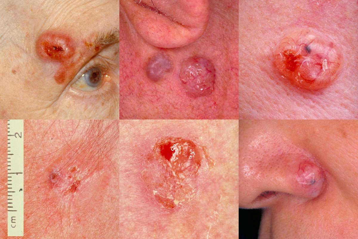
Basal Cell Cancer cancer typically appears as a white, waxy lump or a brown, scaly patch on sun-exposed areas, such as the face and neck. Basal Cell Cancer Treatments include prescription creams or surgery to remove cancer. In some cases, radiation therapy may be required.
 Basal Cell Cancer
Basal Cell Cancer Symptoms
Basal cell carcinomas are usually:
- a single, small, and firm lesion
- dome-shaped and flesh-coloured
- raised at the edges
- pearly or milky white bordered
- covered with tiny blood vessels - easily seen over the top and around the tumour
- may look like a pimple that won’t heal - ulcerated and bleeding at the centre
NODULAR Basal Cell Cancer
Clinically, nodular BCCs comprise 60% of all BCCs. These start off as flat lesions that are best seen when you stretch the skin. Stretching the skin helps you define the edges of the lesion. They slowly grow into small bumps or papules; the most common description of nodular BCC is “pearly papules with a rolled edge.” By a “rolled edge,” we mean that so many of these basal cells start off being dome-shaped, then as they grow laterally, the middle sinks and there is a little ripple all around the edge of the lesion. And on the surface within there will be little tortuous blood vessels. As the lesions grow, they retain the slightly raised border and, even very early on, when the skin is stretched they become bumps. There is a tendency for them to ulcerate in the middle. As they advance, the middle collapses, so if you leave them long enough, these nodular basal cells present as ulcers. In the past, they were called rodent ulcers because they just continue to eat away. They grow laterally and can infiltrate deeply, which is the problem. Most nodular BCCs are on the face and, if left untreated, they are particularly destructive around the nose and the eyes. Usually, nodular BCCs are very well-defined, which is important for treatment: you know where they begin and where they end. This distinguishes them from the ones that are ill-defined. The latter are more difficult to treat.
PIGMENTED Basal Cell Cancer
Pigmented BCCs behave like nodular BCCs, but they have pigment within them that can either be flecks of brown or black scattered through the lesions, or they sometimes can be completely pigmented. This is very important in the differential diagnosis: pigmented BCCs behave like nodular basal cells, but they can be easily confused with melanomas. Conversely, some melanomas do not have a lot of pigment in them. These amelanotic melanomas — which are scary because the risk of missing them is so great — can be a little bit raised, the skin colour is sometimes red and they have flecks of pigment in them. Pigmented BCCs are not that common, but they do occur.
FIBROSING OR SCLEROTIC Basal Cell Cancer
The fibrosing or sclerotic BCCs are usually found on the face. They generally present as what looks like a scar, but without any history of trauma. They have a lot of very dense fibrous stroma, which is what makes them look like scars. Fibrosing BCCs have small, scattered basal cells throughout, making them behave more aggressively. It is more difficult to get rid of them. They are usually flat or slightly depressed, can be yellowish in colour and the surface tends to be smooth and shiny. They are often ill-defined at the borders. Unlike nodular BCC, which generally is soft and gelatinous, fibrosing BCCs are firm, so it is not possible to completely curette them. We can scrape off nodular BCCs because they are much softer than the surrounding skin. Fibrosing BCCs are also more diffuse, which is detectable in the pathology. Therefore, the treatment is different for sclerotic BCCs.
SUPERFICIAL Basal Cell Cancer
Superficial BCCs are thought to be about 15% of BCCs. Because sBCC spreads outward, it was originally thought that it was multifocal cancer, each developing individually. But it is actually all linked together and part of the same single lesion. Superficial BCC occurs as down growths from the undersurface of the epidermis. Superficial BCCs all start off in the epidermis or in the lining of hair follicles and grow down into the dermis. There are not clumps of basal cell in the dermis, as with other BCCs. Superficial BCCs are erythematous, slightly scaly, well-defined patches, most commonly found on the trunk and the limbs, not usually on the face. They are easily confused with a patch of psoriasis or eczema. Psoriasis usually consists of multiple lesions — the same with eczema— and psoriasis tends to be symmetrical. An sBCC is just a lonely little patch of red, well-defined, slightly scaly skin that may not change very much for months or even years. That, in itself, is a clue. If it is just slowly growing, and the patient does not have other, similar lesions, then it might be an sBCC. The features of sBCC are fairly distinctive upon closer inspection. It too has a very fine beading or fine pearly edge — when the skin is stretched — but there might be some fine scaling and some crusting in the middle. As the sBCCs mature, the middle part tends to smooth out and almost look a little bit scared. It becomes a little bit atrophic, thinner, smoother and less red. Superficial Basal Cell Cancer is a commonly misdiagnosed disease; mostly misdiagnosed as psoriasis, discoid eczema or fungus.
FIBROEPITHELIOMA OF PINCUS
Fibroepithelioma of Pincus is rare and different from other BCCs. It tends to be a smooth, elevated little nodule that is sometimes pedunculated. It almost looks like a firm skin tag. It is most often seen on the back or the extremities, but can also be found in the groin or on the sole of the foot. As those are not sun-exposed areas, this disease is probably not sun-related.
You must be logged in to post a comment.